Arthritis of the Shoulder Anatomy
Your shoulder is made up of three bones: your upper arm bone (humerus), your shoulder blade (scapula), and your collarbone (clavicle).The head of your upper arm bone fits into a rounded socket in your shoulder blade. This socket is called the glenoid. A combination of muscles and tendons keeps your arm bone centered in your shoulder socket. These tissues are called the rotator cuff.There are two joints in the shoulder, and both may be affected by arthritis. One joint is located where the clavicle meets the tip of the shoulder blade (acromion). This is called the acromioclavicular (AC) joint.Where the head of the humerus fits into the scapula is called the glenohumeral joint.


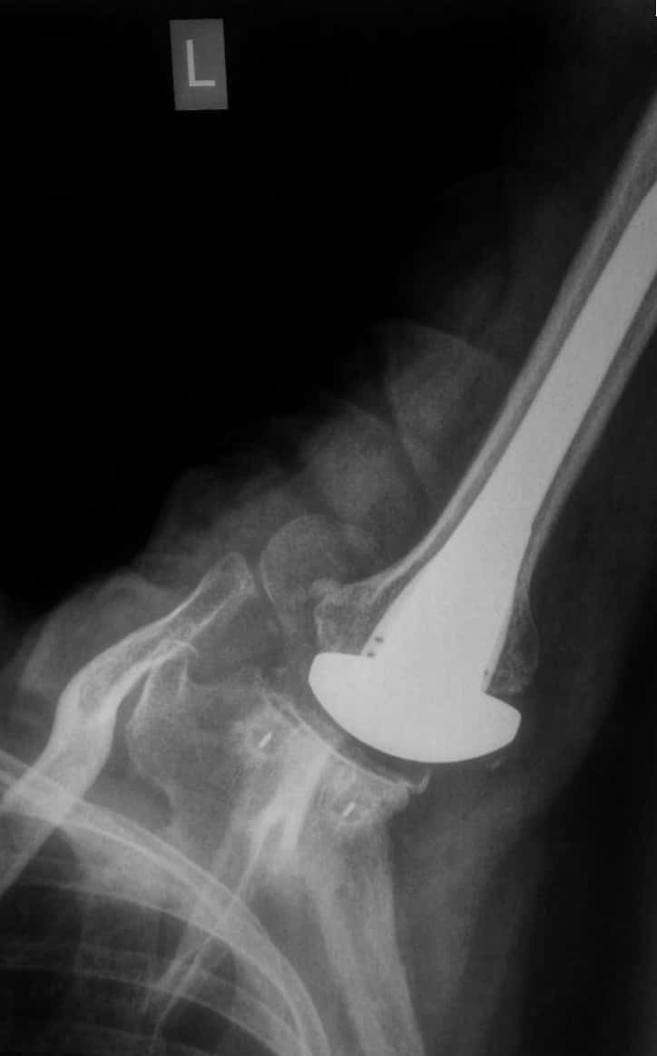
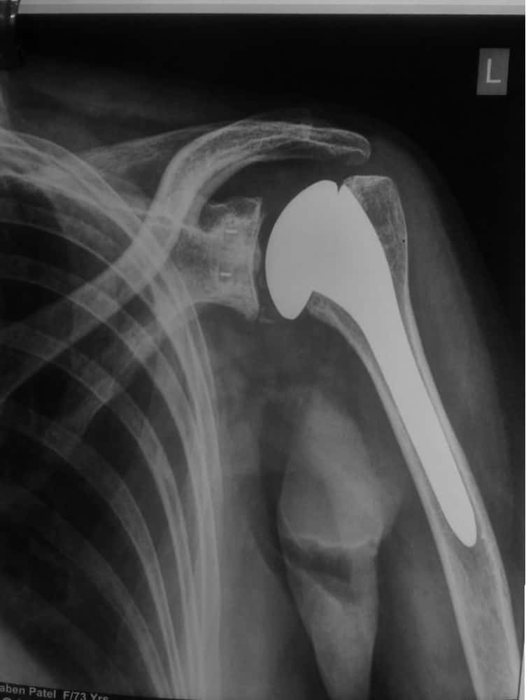
A Typical X-Ray of a well functioning Shoulder Replacement
Osteoarthritis
Also known as “wear-and-tear” arthritis, osteoarthritis is a condition that destroys the smooth outer covering (articular cartilage) of bone. As the cartilage wears away, it becomes frayed and rough, and the protective space between the bones decreases. During movement, the bones of the joint rub against each other, causing pain.
Symptoms
Pain: The most common symptom of arthritis of the shoulder is pain, which is aggravated by activity and progressively worsens.
Limited range of motion: Limited motion is another common symptom. It may become more difficult to lift your arm to comb your hair or reach up to a shelf. You may hear a grinding, clicking, or snapping sound (crepitus) as you move your shoulder.
- Fracture Around The Shoulder
- Shoulder Arthroscopy
- Shoulder Impingement
- Rotator Cuff Tear
- frozen Shoulder
- Shoulder dislocation
- Shoulder Joint Replacement
- Biceps Tendinitis
- Reverse Shoulder Replacement Surgery
- Calcific Tendinitis
- Elbow Replacement
- Elbow Arthroscopy
- Tennis Elbow
- Wrist Fractures
- Wrist Scaphoid Fractures
- Wrist Arthroscopy
- Rheumatoid Wrist
- Carpal Injuries
- Wrist Scaphoid Nonunion
Services
X-Rays
X-rays are imaging tests that create detailed pictures of dense structures, like bone. They can help distinguish among various forms of arthritis.X-rays of an arthritic shoulder will show a narrowing of the joint space, changes in the bone, and the formation of bone spurs (osteophytes).
Treatment
Nonsurgical TreatmentAs with other arthritic conditions, initial treatment of arthritis of the shoulder is nonsurgical. Your doctor may recommend the following treatment options:
Rest or change in activities to avoid provoking pain. You may need to change the way you move your arm to do things.Physical therapy exercises may improve the range of motion in your shoulder.Pain medicationCorticosteroid injections in the shoulder can dramatically reduce the inflammation and pain. However, the effect is often temporary.Moist heatIce your shoulder for 20 to 30 minutes two or three times a day to reduce inflammation and ease pain.
Surgical Treatment
Shoulder joint replacement (arthroplasty).Advanced arthritis of the glenohumeral joint can be treated with shoulder replacement surgery, in which the damaged parts of the shoulder are removed and replaced with artificial components, called prosthesis, just like a knee replacement which is indeed more common and more familiar for the patient!!

Complete wear or loss of normal smooth cartilage over the humeral head (Ball), Damaged cartilage over glenoid (socket), Replaced socket, replaced ballthis is how the replaced shoulder typically looks on an x ray!!
Recovery Surgical treatment of arthritis of the shoulder is generally very effective in reducing pain and restoring motion. Recovery time and rehabilitation plans depend upon the type of surgery performed.
Complications As with all surgeries, there are some risks and possible complications. Potential problems after shoulder surgery include infection, stiffness, excessive bleeding, blood clots, and damage to blood vessels or nerves.
